[headline style=”1″ align=”center” headline_tag=”h2″]
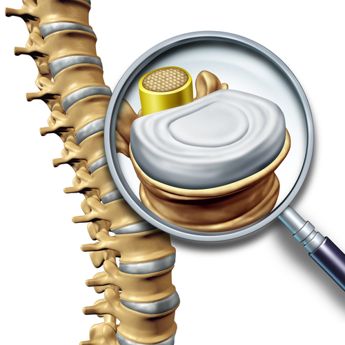
Introduction to Lateral Foraminal Stenosis
[/headline]
Lumbar spinal stenosis is a condition that causes numbness, tingling, weakness, and even pain down one leg or both legs. Often times it is misinterpreted as sciatica and usually impairs those aged 50+.
Treatments for spinal stenosis ranges from flexion biased exercises, core strengthening, mobilization of the spine, and patient education. Surgery is another option if the condition has severely progressed.
In this post, I show you how a 65 YO YO female increased her function with non-traditional physical therapy flexion exercises. Rather, a 3D movement perspective was utilized through treatment of hip and calf mobility.
[headline style=”1″ align=”center” headline_tag=”h2″]
Case History and Examination
[/headline]
A 65 YO female came to PT with a CC of left lateral and posterior thigh and leg pain and distal paresthesia especially when walking greater than 10 minutes. Symptoms were worst later in the day and better in the morning. Sitting alleviated s/s. Patient denied any trauma or any s/s of malignancy.
[headline style=”1″ align=”center” headline_tag=”h2″]
The Examination
[/headline]
Structural Inspection
Forward head and rounded shoulders. Anterior pelvic tilt. Internal rotation of femurs B.
Neuro Examination:
AROM:
Normal spinal motion with no increase in s/s.
Quadrant Test: negative
Accessory:
3/6 lumbar spine
Neuro Examination:
1+ Reflexes on L
4/5 strength of L4 and L5 Myotomes
Light touch sensation: WNL
Deep sensation: Impaired L4 and L5.
Neural Tension Tests Negative
3DMAPS Movement Analysis:
Moderately limited left anterior chain with reduced ankle DF and hip extension.
Moderately limited left same side lateral chain with possibility of hip adductor tightness.
Extremity Evaluation:
Limited ankle DF LR with normal joint mobility indicating calf tightness
Positive Thomas Test LR
2/6 Anterior Glide of Femur
Positive adductor tightness
MMT: Unremarkable
PFT: Unremarkable
[headline style=”1″ align=”center” headline_tag=”h2″]
My Interpretation of Patient’s Examination
[/headline]
Based on the above data, I discerned this patient has s/s that presents with lateral foramina stenosis.
This patient was negative for all neural tension tests, no provocation with piriformis or hamstring lengthening or contraction in SLUMP and ASLR tests, hypo reflexes L>R, myotomal weakness, and loss of deep vibration sensation on L in dermatomal patterns of L4 and L5.
In a perfect scenario, a two stage treadmill test could have been done to rule out vascularity but who has time for that in the clinic?
In this patient’s case, she didn’t have any increase in s/s with spinal movement or with quadrant position indicating possibly that its not so much degenerative changes such as spondylosis. Although I am certain her MRI will show these changes 🙂
Rather, it was something abnormally occurring when walking that was increasing the patient’s signs and symptoms just enough to put pressure on the nerve root.
Even though she probably has degenerative changes of her spine, her stenosis may not have been severe enough to cause symptoms during other movements and positions, just walking.
Clinically, most stenosis patients have pain when standing for too long and performing other activities similiar on their feet. This patient didn’t have this.
It has been my clinical experience you have two categories of stenosis patients and this patient fell into the second one.
The first one is for those that have some serious degenerative changes and compression of the nerve root and have increase in symptoms no matter what they do upright. These patients will often be positive for symptoms with quadrants test or any other motion closing the foramen.
These patients will need modifications to daily activities, traditional flexion biased exercises, and to lay down mid day to allow their disc to imbibe.
The second is those that are older and may have stenotic changes of their spines under imaging but have no s/s with movement of the spine, only increase in symptoms with specific activities. This was the category that my current patient fell in. Only symptoms with walking and not standing or closing of foramen during AROM assessment.
[headline style=”1″ align=”center” headline_tag=”h2″]
Interventions
[/headline]
Based on the above examination I treated the patient with the following exercise and manual therapy strategies below.
My initial thought process was to attack the hip mobility since the patient only had increase in s/s with walking. When the hip is limited in the trail leg of gait further lumbar extension usually occurs as a compensation. This would have increased compression on the nerve root.
Not only was this patient limited in the sagittal plane but also in the frontal plane with adductor tightness. When the adductors are tight they will further limit hip extension in the trail leg in gait since it is going through hip abduction, extension, and internal rotation.
After clearing out the hip, the patient could walk further but was still getting increase in s/s.
Taking another peak at her 3DMAPS Anterior Chain she still had limitations but it did improve.
I then took the path towards going after the posterior calf. After doing some quick ART and 3D stretching to the calf the patient’s anterior chain improved drastically.
Posterior calf tightness does and can limit hip extension. This is one of the many reasons why knowing your biomechanics COLD is crucial for your success as a clinician.
Anyways, enough talking and here are my interventions below:
-
Video 1: Hip extension mobilization with opening of foramen in transverse plane
-
Video 2: Hip abduction mobilization with opening of lumbar foramen in transverse plane
-
Video 3: Hip Abduction (Gait Specific)
-
Video 4: 3D Hip Flexor Stretch
-
Video 5: 3D Calf Stretch
-
Video 6: Anterior Lunge with Arm Driver Ipsilateral Pelvic Motion
-
Video 7: Lunge with Arm Driver Contralateral of Pelvis
[video_player type=”embed” width=”560″ height=”315″ align=”left” margin_top=”0″ margin_bottom=”20″]![]() [/video_player]
[/video_player]
[video_player type=”embed” width=”560″ height=”315″ align=”left” margin_top=”0″ margin_bottom=”20″]![]() [/video_player]
[/video_player]
[headline style=”1″ align=”center” headline_tag=”h2″]
The Outcome
[/headline]
After 8 PT visits this patient was now able to walk symptom free.
The patient’s strength, reflexes, and vibration sense all improved as well.
Not only this, her movement patterns in the anterior and same side lateral chains improved drastically. In retrospect, I wish I would ave taken objective measurements or a video of before and after photos; however, I let results speak for themselves.
[headline style=”1″ align=”center” headline_tag=”h2″]
Concluding Remarks on Spinal Stenosis
[/headline]
Spinal stenosis can be a troublesome condition for both patients and clinicians. By applying traditional physical therapy or, in this case, mobilization of the hips and calf can reduce closure of the lumbar foramen when walking. By classifying your patients in accordance to the two different categories of stenosis the appropriate treatment strategy can be applied.
Have you found something similiar clinically?
P.S. If you are looking to learn more how about this form of treatment you can check out my friends at the Gray Institute by Clicking Here
